Musculoskeletal disease in children and teenagers: Addressing an emerging epidemic
Musculoskeletal conditions represent a common and costly burden to the American health care system.
Musculoskeletal conditions represent a common and costly burden to the American health care system. Roughly 1 of 7 Americans receives care for a musculoskeletal problem,1 and more than $250 billion is spent each year on caring for these patients (Figure 1).
Indeed, more than half of injuries involve the musculoskeletal system.2 Arthritis, osteoporosis, and trauma are the most common problems. Arthritis will affect more than 60 million persons by the year 2020.3 One of every 2 women and 1 of every 8 men older than 50 years will sustain a fracture related to osteoporosis in their lifetime.1
Overall, musculoskeletal problems have a significant impact on the health care system and quality of life-not only in adults but also in children and teenagers. Although these problems often are discussed as issues that affect older persons, in many cases they begin at a young age and progress to end-stage disease during adulthood. Given these facts, developing musculoskeletal education programs for children and teenagers is an important step toward improving injury prevention to preserve quality of life and to decrease the cost of care.
In this article, we provide a review of trends in musculoskeletal trauma, arthritis, and osteoporosis in children and teenagers. We also discuss the importance of early education in injury prevention.
MUSCULOSKELETAL CONDITION
Trauma
Pediatric trauma remains a significant cause of morbidity and mortality in children and teenagers. It affects 1 of every 3 to 4 children, and up to 22 million children sustain injuries each year.4
Musculoskeletal injuries most often result from road accidents and recreational activities. Traumatic injuries, especially those involving motor vehicles, is the leading cause of fatalities among children and teenagers.2,4,5 Road injuries also maim millions in this age group each year. In addition, falls and sports injuries constitute the largest group of injuries resulting in hospitalization or emergency department visits.2
Recreational activities account for a significant number of injuries in children and teenagers (Figure 2 and Figure 3). During the past 2 decades, high school sports participation has increased significantly and, as a result, overuse injuries are becoming more frequent. About 10% of children participating in organized sports will be injured each year.6
More than 2 million children aged 5 to 14 years are treated medically each year for musculoskeletal injuries resulting from participation in the following 8 common types of recreational act


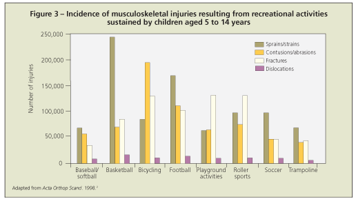
ivities (in order of decreasing incidence of musculoskeletal injury): bicycling, basketball, football, roller sports, playground activities, baseball, soccer, and trampoline.6 With current safety guidelines and protective equipment, many of these injuries are preventable.6
Children and teenagers sustain more injuries in free play than in organized sports.7 The two most common causes of sports fatalities are head injuries and cardiac events.6 Heat stroke is the third most common cause of exercise-related death among high school athletes.8 The highest rate of catastrophic injuries is seen in gymnastics.8
Bicycle riding injuries-including contusions, abrasions, sprains, strains, fractures, and dislocations-account for the highest number of recreational injuries sustained by patients aged 5 to 14 years.6 In contact sports, sprains, strains, and contusions are the most common musculoskeletal injuries.9
Playgrounds are the second most common location where unintentional injuries occur in children. Falls are the most common mechanism of injury on public and private playgrounds.10 Fractures are the most commonly reported injury with falls.6
The costs attributable to pediatric trauma are significant. The estimated cost of unintentional childhood injuries is $7.2 billion.2
Perhaps more important than the financial costs of pediatric trauma is the associated physical and emotional stress. Many children are left with permanent physical, psychological, and socioeconomic impairments, which are difficult to measure in dollars. Persistent pain and disability affect not only the capacity to do work but also the ability to perform activities of daily living. In children and teenagers, posttraumatic chronic pain may be crippling and significantly interfere with school attendance and job placement.
Wesson and associates11 studied the impact of musculoskeletal injuries on children using the Injury Severity Score (ISS); 71% of children with major injuries (ISS higher than 16) and 54% of those with minor injuries (ISS lower than 16) had some residual physical impairment 12 months after injury, compared with 0% of controls.11 The same study showed a sharp rise in the rate of behavioral disturbances in children with major injuries, and they tended to persist indefinitely in those with physical impairment. A decrease in academic performance was noted in patients who had major injury both with and without head injury. Fewer than two-thirds of the patients' families returned to a normal life, compared with 100% of control families.11 These findings underscore the significant psychological and socioeconomic impact of trauma on the pediatric population and their families.
Developing programs that stress preventive measures may be the best strategy to decrease the incidence of pediatric musculoskeletal trauma.12 Efforts to improve road safety should include education about motor vehicles, traffic laws, the use of passive restraints (seat belts, car seats), and the dangers associated with driving under the influence of drugs or alcohol. The implementation of mandatory seat belt laws has significantly reduced the number of injuries associated with motor vehicles.2
Prevention efforts in recreational activities should focus on teaching proper techniques and encouraging the use of protective gear. Implementation of these strategies has clearly reduced the number of injuries in football, baseball, and bicycling. In football, for example, the development of a more protective helmet and safer tackling techniques significantly reduced the number of head and neck injuries.6
In baseball, repetitive and improper throwing techniques (sidearm throwing, curve balls) account for most elbow injuries in children and teenagers.6 Instructing coaches about proper pitching techniques and implementing pitch counts has reduced the number of elbow injuries.6 In addition, the use of breakaway bases has prevented more than 1 million baseball-related injuries each year.13 For bicyclists, implementation of mandatory helmet laws has resulted in a decline in severe head injuries.14
General strategies used to prevent injuries related to recreational activities include the following:
•Performing a thorough preparticipation history and physical examination.
•Gaining an understanding of the participation guidelines put forth for certain medical conditions, as defined by the American Academy of Pediatrics.
•Encouraging the use of sports-specific protective equipment.
•Following participation guidelines defined by age, weight, and ability.
•Developing realistic expectations in coaches and parents.
Overall, pediatric trauma remains a significant socioeconomic burden. National efforts aimed at injury prevention-in addition to continuing advances in medical care-are essential to make a significant impact on reducing the rate of musculoskeletal injuries in children and teenagers.
Arthritis
This condition is the leading cause of disability in the United States. More specifically, osteoarthritis (OA)-one of the most common progressive musculoskeletal diseases in adults-has a significant physical, psychological, and economic impact on society. Adults with OA have direct health care costs nearly double those of age-matched controls.15 In 2003, the direct medical expenditures for arthritis and other rheumatologic conditions was more than $80 billion.2
Although the prevalence of OA in children and teenagers is low, it increases steadily with age. In fact, the prevalence of arthritis is 8% in persons 18 to 44 years old and rises to 50% in persons older than 65 years.16
Even though OA is not as widespread in younger persons, certain factors place them at greater risk for this condition early in life. They include obesity, damage to articular cartilage, malalignment of the lower extremities, and irreparable damage to the meniscus or labrum. Certain pediatric orthopedic disorders (eg, hip dysplasia, slipped capital femoral epiphysis [SCFE], and adolescent Blount disease) also increase the chance of early OA.
Joint injury significantly increases a person's risk of OA. This factor is becoming a more significant issue with the rise in injuries in the pediatric population.
For example, irreparable meniscal tears and anterior cruciate ligament (ACL) injuries predispose children and teenagers to degenerative joint disease.17-19 Lohmander and colleagues20 evaluated female soccer players and found that previous knee injury was correlated with subsequent development of OA in the injured joint. Gelber and coworkers21 monitored a cohort of more than 1300 young patients (average age, 22 years) and demonstrated that persons with knee injuries at baseline evaluation are at significantly increased risk for OA.
Although no studies have confirmed that ACL reconstruction lowers the risk of OA, recent studies suggest that specific neuromuscular training can be used to prevent ACL injuries.22,23 Even though successful management of sports-related injuries allows athletes to return to play, effective early management does not preclude the increased probability of OA.24 In fact, even if no injury occurs, vigorous participation in recreational activities as a child or teenager may increase the probability of OA.24
During the past 25 years, the prevalence of obesity in the United States has quadrupled in children. About 30% of children and teenagers are overweight or at increased risk for becoming overweight.25 In addition, adolescent obesity is predictive of obesity during adulthood. Recently, more attention has been directed toward the social, medical, and economic effects of obesity. Elevated body mass index (BMI) is a major risk factor for the development of degenerative joint disease.26 In fact, an elevated BMI value in persons aged 20 to 29 years increases the risk of OA by more than 3-fold.26
The increased weight associated with obesity imparts more cumulative stress to the knee joint. It also increases the risk of varus or valgus angular deformity. This acquired malalignment coupled with an increase in force across the joint may contribute to early degenerative joint disease of the knee.27-30
Increased body weight also places children at greater risk for SCFE and Blount disease. Both conditions may lead to abnormal joint function and cause early OA.31
Careful clinical evaluation of body weight, lower extremity alignment, the hips, and the knees is required in all children and teenagers. Early diagnosis and intervention often reduces the risk that these disorders will progress to early degenerative joint disease. A recent systematic review of the literature concluded-with cautious optimism-that the combination of nutritional education, physical activity, and family support are important steps in preventing childhood obesity.32-36
Overall, the development of OA is multifactorial. Physicians should be aware of the controllable risk factors and educate patients about them to establish good bone health at an early age.
Osteoporosis
This is a significant musculoskeletal health issue. By 2020, half of Americans older than 50 years are expected to have or to be at risk for osteoporosis. The cost of managing osteoporotic fractures exceeded $15 billion in 2002, and this figure will double or triple in the coming decades.1
Osteoporosis is rare in children and teenagers. However, 40% of bone mass accumulates during adolescence. The bone mineral density (BMD) established during this period helps determine bone health and define the risk of osteoporosis in adulthood.37,38
Osteoporosis in children and teenagers develops as a primary condition, such as juvenile idiopathic osteoporosis or osteogenesis imperfecta. It also develops secondary to chronic diseases, especially those that require long-term corticosteroid therapy.
Data currently are insufficient to formally define osteoporosis in children and teenagers. The guidelines based on dual-energy x-ray absorptiometry (DEXA) scans used for adults, including T-scores, are not applicable to this age group.
As an alternative, Z-scores are used to help define the extent of osteopenia in young patients.39 In those younger than 20 years, the terminology "low bone density for chronological age" may be used if the Z-score is lower than 22.40 Although Z-score values lower than 22 generally are considered a serious warning, most bone specialists make a diagnosis of osteoporosis in children and adolescents only in the presence of low BMD and at least 1 fragility fracture.41
Low calcium intake and inadequate physical activity in young persons increase their risk of developing osteoporosis. Anderson and associates42 surveyed adolescents about their knowledge and beliefs about bone health and found that most teens do not know that weight-bearing exercise reduces the risk of osteoporosis. In fact, more than half of the adolescents rated their physical activity level as low or moderate. The adolescents also had difficulty in identifying foods that are high in calcium, and 58% of them had low calcium intake.
Many of the participants had risk factors for the development of osteoporosis. For example, 25% of the adolescents defined themselves as smokers, and cigarette smoking has a negative effect on BMD. In a meta-analysis examining the link between smoking and osteoporosis, cigarette smoking was reported as an independent risk factor for lower bone density and increased risk of fracture.43
Caffeine, phosphorus, and carbonation of soda are among the dietary factors related to bone health that interfere with calcium absorption, which leads to low bone mass.1 Adequate calcium and vitamin D intake during childhood leads to increased bone mass (Table 1 and Table 2).44-46 Kalkwarf and coworkers47 analyzed data from the National Health and Nutrition Examination Survey and found that women who report having drunk milk regularly during childhood have higher hip bone density and a lower fracture risk than those who do not.
Weight-bearing exercise also increases BMD.48,49 In one study, children randomized to participation in a high-impact exercise program had higher hip BMD density than age-matched controls. This effect persisted when the children underwent repeated DEXA scanning 7 months after stopping the exercise program.49 Similar gains in BMD were seen in the hips and spines of children who engaged in a 2-year school-based jumping program.50
Overall, prevention of osteoporosis begins in childhood. Clinicians should counsel young patients and their families that adequate nutrition and weight-bearing activity are necessary for good bone health.
EARLY EDUCATION IS IMPORTANT
The average American consumes levels of calcium far below the amount recommended for optimal bone health, according to the Surgeon General.1 In addition, even though genetic factors play a significant role in determining bone mass, controllable lifestyle factors (eg, diet and physical activity) can mean the difference between a frail skeleton and a strong one. Physical fitness helps decrease the risk of obesity and of low BMD.24 Efforts to maintain bone health early in life can result in a strong skeleton later in life. Teaching children and teenagers about lifestyles that promote good bone health is an essential component of addressing this major public health problem.
The Bone & Joint Decade 2000-2010 has developed several programs to educate the public about bone health. For example, the Protect Your Bones program, which started at the Cleveland Clinic, provides an opportunity to teach young persons about making appropriate decisions each day to promote good bone health.
The program is divided into 3 parts: assessment of knowledge, discussion of topics, and question and answers. A prelecture quiz determines the participant's baseline knowledge of the lecture topics. The interactive lecture then focuses on trauma, arthritis, and osteoporosis. Within each section, the topic is defined, statistics are discussed to emphasize the relevance of the problem, and then instructions are given on how to take steps to preserve bone health with respect to the topic being discussed. After questions are answered, a postlecture quiz is given to assess what the students have learned from the lecture.
Implementation of the Protect Your Bones program has resulted in significant improvement in how teenagers understand musculoskeletal disease. Based on quiz results, the students statistically improved their understanding of trauma by 50%, arthritis by 30%, and osteoporosis by 15%. More important, more than 80% of the students strongly agreed that they learned something from the lecture, that the information was worth sharing with their families, and that the program should be continued for other students.
Several organizations, such as the NIH and the American Dairy Association, are implementing similar programs to improve understanding of musculoskeletal disease among children and teenagers. Regardless of which program appeals to a physician, everyone has a role to play in improving and promoting bone health. Physicians' participation is essential to broaden the public's understanding of these issues. More research also is needed to further define the efficacy of education programs on preventing musculoskeletal injuries, arthritis, and osteoporosis. More information on musculoskeletal education programs is available on the Web sites for the US Surgeon General (www.surgeongeneral.gov) and for the Bone & Joint Decade Web site (www.usbjd.org).
References:
References1. Surgeon General of the United States. Bone Health and Osteoporosis: a Report of the Surgeon General. Washington, DC: US Department of Health and Human Services; 2004.
2. The Bone & Joint Decade 2000-2010 for prevention and treatment of musculoskeletal disorders. Acta Orthop Scand Suppl. 1998;69:S1-S20.
3. Praemer A, Furner S, Rice D. Musculoskeletal Conditions in the United States. Rosemont, IL: American Academy of Orthopaedic Surgeons (AAOS); 1999.
4. Galano GJ, Vitale MA, Kessler MW, et al. The most frequent traumatic orthopaedic injuries from a national pediatric inpatient population. J Pediatr Orthop. 2005;25:39-44.
5. Hicks BA, Morris JA Jr, Bass SM, et al. Alcohol and the adolescent trauma population. J Pediatr Surg. 1990;25: 944-949.
6. Purvis JM, Burke RG. Recreational injuries in children: incidence and prevention. J Am Acad Orthop Surg. 2001;9:365-374.
7. Biermann JS, Micheli LJ, Ogden JA, Ireland ML. Injuries and conditions in children and adolescents: which is safer? Organized sports or free play? AAOS Annual Meeting; February 28-March 4, 2001; San Francisco.
8. Sullivan J, Anderson S, eds. Care of the Young Athlete. Rosemont, IL: AAOS and AAP; 2000.
9. Beiner JM, Jokl P. Muscle contusion injuries: current treatment options. J Am Acad Orthop Surg. 2001;9:227-237.
10. Tinsworth DK, McDonald JE. Special Study: Injuries and Deaths Associated With Children's Playground Equipment. Washington, DC: US Consumer Product Safety Commission; April 2001.
11. Wesson DE, Scorpio RJ, Spence LJ, et al. The physical, psychological, and socioeconomic costs of pediatric trauma. J Trauma. 1992;33:252-257.
12. Patterson MM. Prevention: the only cure for pediatric trauma. Orthop Nurs. 1999;18:16-20.
13. Janda DH, Wojtys EM, Hankin FM, et al. A three-phase analysis of the prevention of recreational softball injuries. Am J Sports Med. 1990;18:632-635.
14. Mock CN, Maier RV, Boyle E, et al. Injury prevention strategies to promote helmet use decrease severe head injuries at a level I trauma center. J Trauma. 1995;39:29-35.
15. Mapel DW, Shainline M, Paez K, Gunter M. Hospital, pharmacy, and outpatient costs for osteoarthritis and chronic back pain. J Rheumatol. 2004;31:573-583.
16. Prevalence of doctor-diagnosed arthritis and arthritis-attributable activity limitation-United States, 2003-2005 [published corrections appear in MMWR. 2006;55:1129 and 2007;56:55]. MMWR. 2006;55:1089-1092.
17. Englund M, Roos EM, Lohmander LS. Impact of type of meniscal tear on radiographic and symptomatic knee osteoarthritis: a sixteen-year followup of meniscectomy with matched controls. Arthritis Rheum. 2003;48:2178-2187.
18. Roos H, Laurén M, Adalberth T, et al. Knee osteoarthritis after meniscectomy: prevalence of radiographic changes after twenty-one years, compared with matched controls. Arthritis Rheum. 1998;41:687-693.
19. von Porat A, Roos EM, Roos H. High prevalence of osteoarthritis 14 years after an anterior cruciate ligament tear in male soccer players: a study of radiographic and patient relevant outcomes. Ann Rheum Dis. 2004;63:269-273.
20. Lohmander LS,Ostenberg A, Englund M, Roos H. High prevalence of knee osteoarthritis, pain, and functional limitations in female soccer players twelve years after anterior cruciate ligament injury. Arthritis Rheum. 2004;50:3145-3152.
21. Gelber AC, Hochberg MC, Mead LA, et al. Joint injury in young adults and risk for subsequent knee and hip osteoarthritis. Ann Intern Med. 2000;133:321-328.
22. Hewett TE, Ford KR, Myer GD. Anterior cruciate ligament injuries in female athletes, part 2: a meta-analysis of neuromuscular interventions aimed at injury prevention. Am J Sports Med. 2006;34:490-498.
23. Mandelbaum BR, Silvers HJ, Watanabe DS, et al. Effectiveness of a neuromuscular and proprioceptive training program in preventing anterior cruciate ligament injuries in female athletes: 2-year follow-up. Am J Sports Med. 2005;33:1003-1010.
24. Garrick JG, Requa RK. Sports fitness activities: the negative consequences. J Am Acad Orthop Surg. 2003;11:439-443.
25. Ogden CL, Flegal KM, Carroll MD, Johnson CL. Prevalence and trends in overweight among US children and adolescents, 1999-2000. JAMA. 2002;288:1728-1732.
26. Gelber AC, Hochberg MC, Mead LA, et al. Body mass index in young men and the risk of subsequent knee and hip osteoarthritis. Am J Med. 1999;107:542-548.
27. Beskin JL, Burke SW, Johnston CE 2nd, Roberts JM. Clinical basis for a mechanical etiology in adolescent Blount's disease. Orthopedics. 1986;9:365-370.
28. Dietz WH Jr, Gross WL, Kirkpatrick JA Jr. Blount disease (tibia vara): another skeletal disorder associated with childhood obesity. J Pediatr. 1982;101:735-737.
29. Gushue D, Houck J, Lerner A. Effects of childhood obesity on three-dimensional knee joint biomechanics during walking. J Pediatr Orthop. 2005;25:763-768.
30. Henderson RC. Tibia vara: a complication of adolescent obesity. J Pediatr. 1992;121:482-486.
31. Morrissy RT. Slipped capital femoral epiphysis. In Morrissy RT, ed. Lovell and Winter's Pediatric Orthopaedics. 3rd ed. Philadelphia: JB Lippincott; 1990:902.
32. Bautista-Castaño I, Doreste J, Serra-Majem L. Effectiveness of interventions in the prevention of childhood obesity. Eur J Epidemiol. 2004;19:617-622.
33. Clark H, Goyder E, Bissell P, et al. How do parents' child-feeding behaviours influence child weight? Implications for childhood obesity policy. J Public Health (Oxford). 2007;29:132-141.
34. Felson DT, Zhang Y, Anthony JM, et al. Weight loss reduces the risk for symptomatic knee osteoarthritis in women. The Framingham Study. Ann Intern Med. 1992; 116:535-539.
35. Golan M, Weizman A, Apter A, Fainaru M. Parents as the exclusive agents of change in the treatment of childhood obesity. Am J Clin Nutr. 1998;67:1130-1135.
36. Sothern MS. Exercise as a modality in the treatment of childhood obesity. Pediatr Clin North Am. 2001;48:995-1015.
37. Saggese G, Baroncelli GI, Bertelloni S. Osteoporosis in children and adolescents: diagnosis, risk factors, and prevention. J Pediatr Endocrinol Metab. 2001;14:833-859.
38. Theintz G, Buchs B, Rizzoli R, et al. Longitudinal monitoring of bone mass accumulation in healthy adolescents: evidence for a marked reduction after 16 years of age at the levels of lumbar spine and femoral neck in female subjects. J Clin Endocrinol Metab. 1992;75:1060-1065.
39. Kalkwarf HJ, Zemel BS, Gilsanz V, et al. The bone mineral density in childhood study: bone mineral content and density according to age, sex, and race. J Clin Endocrinol Metab. 2007;92:2087-2099.
40. Baroncelli GI, Bertelloni S, Sodini F, Saggese G. Osteoporosis in children and adolescents: etiology and management. Paediatr Drugs. 2005;7:295-323.
41. Bianchi ML. Osteoporosis in children and adolescents. Bone. 2007;41:486-495.
42. Anderson KD, Chad KE, Spink KS. Osteoporosis knowledge, beliefs, and practices among adolescent females. J Adolesc Health. 2005;36:305-312.
43. Ward KD, Klesges RC. A meta-analysis of the effects of cigarette smoking on bone mineral density. Calcif Tissue Int. 2001;68:259-270.
44. Hoppe C, Mølgaard C, Michaelsen KF. Bone size and bone mass in 10-year-old Danish children: effect of current diet. Osteoporos Int. 2000;11:1024-1030.
45. Mølgaard C, Thomsen BL, Michaelsen KF. The influence of calcium intake and physical activity on bone mineral content and bone size in healthy children and adolescents. Osteoporos Int. 2001;12:887-894.
46. Ruiz JC, Mandel C, Garabedian M. Influence of spontaneous calcium intake and physical exercise on the vertebral and femoral bone mineral density of children and adolescents. J Bone Miner Res. 1995;10:675-682.
47. Kalkwarf HJ, Khoury JC, Lanphear BP. Milk intake during childhood and adolescence, adult bone density, and osteoporotic fractures in US women. Am J Clin Nutr. 2003;77:257-265.
48. Fuchs R, Bauer J, Snow C. Jumping improves hip and lumbar spine bone mass in prepubescent children: a randomized controlled trial. J Bone Miner Res. 2001;16:148-156.
49. Fuchs RK, Snow CM. Gains in hip bone mass from high-impact training are maintained: a randomized controlled trial in children. J Pediatr. 2002;141:357-362.
50. MacKelvie KJ, Khan KM, Petit MA, et al. A school-based exercise intervention elicits substantial bone health benefits: a 2-year randomized controlled trial in girls. Pediatric. 2003;112(6, pt 1):e447.


