Osteoarthritis: A Photo Essay
Osteoarthritis is failure of the synovial joint, as shown in these images. Joint pain is the main clinical feature, as well as joint stiffness and deformity.
In a knee with advanced osteoarthritis, large ulcerated areas of articular cartilage are present in the intercondylar region, on the femoral condyles, and on the patella. Articular cartilage involvement is prominent in OA, but OA is not merely a cartilage disease.


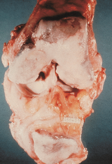
Photo courtesy of Kenneth D. Brandt, MD
Click here for the next image
Microcracks in subchondral bone of an osteoarthritic knee are seen here. They extend into the overlying zone of calcified cartilage and are a manifestation of damage of the subchondral plate.
Photo courtesy of D. B. Burr, PhD
This photograph shows fibrillation of articular cartilage in osteoarthritis. The surface is disrupted, and vertical clefts extend into the depths. When horizontal tears running near the base join the vertical fissures, fragmentation of the joint surface results.


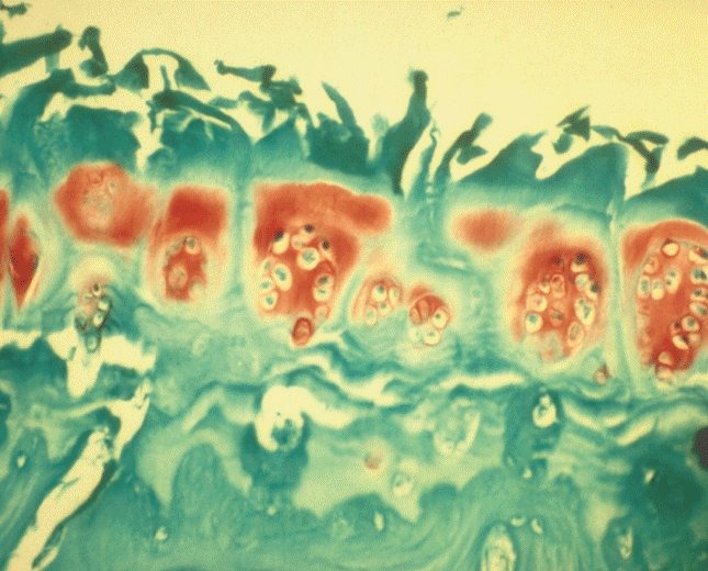
Photo courtesy of Kenneth D. Brandt, MD
Osteoarthritis of the first metatarsophalangeal joint may lead to hallux valgus deformity, with inflammation of the overlying bursa (bunion).


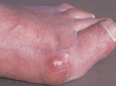
Photo courtesy of Kenneth D. Brandt, MD
Muscle weakness, especially in the quadriceps, may be both a cause and an effect of osteoarthritis of the knee. Bilateral tricompartmental knee OA is shown in this x-ray film, more severe on the right, with subchondral sclerosis. Varus deformity also is noted.


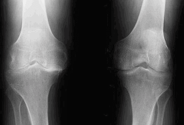
Image courtesy of Christopher R. Phillips, MD and Richard D. Brasington Jr, MD
Click here for the next image
In a patient who has osteoarthritis of the hand, severe first carpometacarpal OA with radial subluxation of the first metacarpal is seen. Mild OA is seen at the proximal interphalangeal joints and more severe changes at the distal interphalangeal joints.


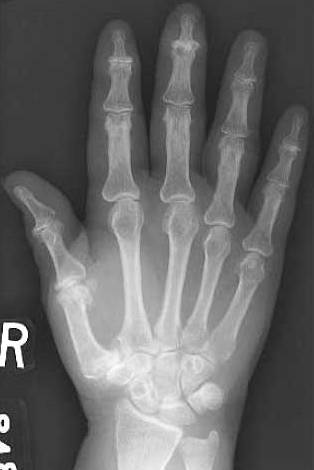
Image courtesy of Christopher R. Phillips, MD and Richard D. Brasington Jr, MD
Click here for the next image
Here, severe osteoarthritis of the hip with subchondral cystic and sclerotic changes is evident in the right hip. There is moderate left hip OA, with superior more than medial joint-space narrowing, as is typical for primary OA. Pain with hip OA usually localizes to the groin.


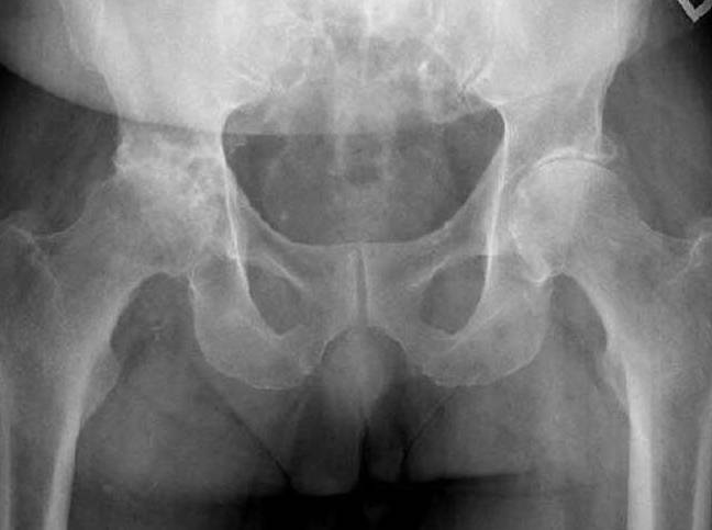
Image courtesy of Christopher R. Phillips, MD and Richard D. Brasington Jr, MD


