Doomed Arthritic Knees Rotate More and Never Rest
New research has examined 3D motion and muscle activity in knee osteoarthritis, and correlated gait with pain. Result: a better description of knees destined for replacement.
“Something in the way she moves.” The Beatles certainly weren’t thinking of an older, overweight woman with bad knees when they sang those words, but they do express the focus of many experts in the biomechanics of knee osteoarthritis (OA). Exactly what is it about that most ordinary daily activity, walking, that impels some people with knee osteoarthritis (most of them heavier women) toward knee replacement?


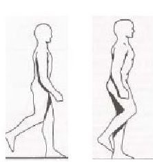
Studies thus far have drawn attention to one factor--differences in the knee adduction moment (KAM), the rotational force or torque exerted by the inward motion of the bending knee during the gait. KAM must play an important role in knee osteoarthritis (OA), because it is the primary determinant of load on the medial compartment of the knee joint, Stanford University professor of engineering and orthopaedics Thomas Andriacchi PhD (a pioneer in OA biomechanics) pointed out a decade ago.
But it’s not the whole story. Research described at the 2013 Osteoarthritis Research Society International conference provides a much clearer image of what goes wrong with a knee that is striding (or limping) toward replacement.
• The muscles supporting doomed knees seem unable to rest. This is one outcome from a triad of studies by a team at Dalhousie University in Halifax, Nova Scotia, who studied 50 patients with moderate medial compartment knee OA during “self-selected walking,” using the standard biomechanics equipment (a force plate in the floor and video images backed by computerized pattern recognition techniques). Among the many factors they assessed, Cheryl Hubley-Kozey PhD, a professor in the Schools of Physiotherapy and Biomedical Engineering, and her coworkers used surface electromyography to measure muscle activation. Five to eight years later, they telephoned the same group to ask about their progress.
Exactly half had undergone total knee replacement (TKR). Despite similar muscle strength, walking velocity, structural impairment, and symptoms on the WOMAC (Western Ontario and McMaster Universities Arthritis Index) at baseline, the group that went on to surgery initially showed consistently higher quadriceps, hamstring, and gastrocnemius activity throughout the gait cycle, Hubley-Kozey reported. This implied higher muscle co-activation (several muscles firing at once), with greater likelihood of muscle fatigue and increased compressive loading exposure on the knee.
• At-risk knees with OA rotate abnormally throughout the entire gait. To date, biomechanics research on knee OA has primarily focused on assessing frontal mechanics. The Dalhousie team analyzed gait as a 3D phenomenon, revealing a stiff gait that reduced the ability of patients destined for knee surgery to “unload” their knee during each step. In biomechanical terms, the only significant 3D gait difference, reported doctoral student Gillian Hatfield, was that people who later chose surgery had, years earlier, shown more external rotation during the stance portion of their gait. However, altered rotational movements were in effect throughout the entire gait, not merely while landing or pushing off, Hatfield added. Consistent with previous reports of increased structural damage, over the entire gait cycle KAM was also greater among knees destined for replacement.
• People with painful knees have a less consistent gait. Few biomechanical studies of knee OA have included symptoms as a variable, and the Dalhousie team addressed this as well. Studies headed by biomechanics professor Janie Astephen-Wilson PhD assessed the same general gait measurements with pain as a variable, by comparing a different set of subjects: 37 symptomatic individuals diagnosed with moderate knee OA and 27 asymptomatic subjects (who all had radiographic evidence of OA in their knees but no pain).
Besides having greater KAM, consistent with earlier research, the symptomatic subjects had systematically lower angles of knee flexion during their gait, and the internal torque measurements on their knees were less constant than those in pain-free subjects. The fact that symptomatic patients had higher KAM may signify that this greater rotational force as their knee bends during a stride may be a response to symptoms, rather than a factor contributing to the structural progress of OA before the onset of the symptoms, she concluded.
Interesting, certainly, but what do the biomechanics of knee OA imply for prevention or treatment? Other research is leading to some practical suggestions.
• Braces and insoles that correct misalignments may prevent progression. Data from 407 subjects in the Dutch PROOF (Prevention of knee OA in Overweight Females) study show a varus (bowleg or < 178 degrees) malalignment in 40% of all knees without OA. This deformity had a significant correlation with the onset of both clinical and structural knee OA in some of these overweight and obese women. (A previous study has shown knock-knees or valgus malalignment also associated with onset and progression of knee OA.) Correcting alignment non-surgically may be a strategy for prevention, concluded Jos Runhaar, a doctoral student at Erasmus Medical Center in Rotterdam, during his presentation.
• The Alexander Technique may help to reduce knee pain resulting from overactive muscles. In a pilot study from the University of Manchester and University College London involving 11 patients with knee OA, 20 sessions of training with the Alexander Technique significantly reduced medial co-contraction between the quadriceps and hamstrings, as measured by electromyography during gait analysis studies. It also significantly improved WOMAC pain scores.
• Specialized footwear can reduce loading on the knee. A pilot study, not presented at the conference but published in Arthritis & Rheumatism two days after it ended, has found that footwear with a flexible sole can substantially reduce the KAM and, by implication, loading on the medial compartment of the knee. Subjects who wore custom “mobility shoes” had an 18% reduction in KAM within 24 weeks, comparable to that produced by barefoot walking. Six months later they even showed reductions in KAM when walking in their own shoes. The custom shoes are sold online at drcomfort.com.


