Identifying and Managing Scapular Problems in Overhead Athletes
The shoulder joint and scapula are inextricably linked-what affects the scapula will affect the shoulder, and vice versa. The scapula houses the glenoid, the bony socket that articulates with the humeral head; by virtue of the scapula's intimate association with shoulder joint motion, abnormalities of the scapula may cause and indicate intrinsic shoulder pathology.
ABSTRACT: Scapular problems in overhead athletes, such as baseball throwers and swimmers, often are responsible for shoulder pathology. Most problems involve dyskinesis, or abnormal motion. In dyskinesis, the scapula may migrate proximally or distally and medially or laterally; protraction is the most common dyskinetic pattern in athletes. Disruption in the kinetic chain may result in overuse and inhibition of periscapular muscles. Physical examination of the athlete's shoulder should include inspection of his or her back to detect dysfunction. A complete biomechanical evaluation should be conducted. Scapular protraction should be mitigated with performance of retraction exercises. All overhead athletes should be encouraged to engage in a dedicated rotator cuff strengthening regimen. (J Musculoskel Med. 2011;28:465-471)
The shoulder joint and scapula are inextricably linked-what affects the scapula will affect the shoulder, and vice versa. The scapula houses the glenoid, the bony socket that articulates with the humeral head; by virtue of the scapula's intimate association with shoulder joint motion, abnormalities of the scapula may cause and indicate intrinsic shoulder pathology. Scapular problems are common in overhead athletes, such as baseball throwers and swimmers. Most of these problems involve dyskinesis, or abnormal motion.
Disorders of the scapula in overhead athletes often go unrecognized; therefore, a shoulder examination is not complete without examination of the posterior shoulder, including the scapula. Effective evaluation of scapular problems in overhead athletes begins with a comprehensive history and physical examination and leads to an appropriate choice of treatment. In most cases of scapular dyskinesis, early diagnosis leads to resolution of symptoms without surgery.
In this article, we discuss disorders of the scapula in overhead athletes, including scapular anatomy, the biomechanics of scapular dyskinesis and the role of the kinetic chain, diagnosis of scapular problems, and the approaches to treatment. Special attention is paid to the role of the scapula as a source of shoulder pain.
ANATOMY
The scapula, a flat bone, has many important functions. They include the following:
•Serves as the origin of virtually every muscle about the shoulder, including the rotator cuff, deltoid, biceps, and triceps.
•Contains the acromion, the anterior scapular projection that is implicated in rotator cuff impingement (abutment, with forward arm flexion, of the acromial process and the supraspinatus tendon). The scapular spine serves as a demarcation between the supraspinatus and the infraspinatus; it is more grossly evident in the presence of significant rotator cuff atrophy.
•Contains the glenoid, the "other end" of the shoulder articulation. With arm motion, the scapula (and glenoid) must "follow" the humeral head to maintain optimal shoulder stability and function by maintaining the physiological "instant center of rotation."1 The glenoid-humerus relationship has been described as a seal balancing a ball on its nose. If the seal cannot follow the ball adequately, it falls off. Similarly, if the scapula does not follow the humeral head properly, glenohumeral instability can result as the humeral head "falls off" the glenoid.
The scapula rests on the ellipsoid thorax and is freely mobile-it has no direct connection to the rib cage. This well-vascularized bone is suspended from the clavicle by the strong coracoclavicular ligaments; it joins the clavicle at the acromioclavicular joint and the humerus at the glenohumeral capsule. Therefore, compromise to these soft tissue connections (acromioclavicular separation) may alter the stability or position of the scapula.
The scapula serves as an attachment to a large amount of muscles. Therefore, when the scapula becomes unstable, it severely disrupts normal shoulder function. It creates an unstable platform from which certain muscles (eg, the rotator cuff) function. It also disrupts the length-tension relationship of several muscles, which causes shoulder muscle weakness, leading to glenohumeral instability.
BIOMECHANICS
Scapular dyskinesis
In scapular dyskinesis, the rhythmic, smooth synchrony among the scapula, thorax, and humerus is disturbed.2 The position of the scapula at rest is also noted to be asymmetrical to that in the contralateral, asymptomatic shoulder.
The scapula rotates about the thorax in 3 axes: abduction/adduction (coronal plane); forward, or anterior, and backward, or posterior, tilting (sagittal plane); and internal rotation/external rotation (axial plane). In addition, the scapula may migrate proximally (elevation) or distally (depression) and medially (retraction) or laterally (protraction). The most frequently recognized dyskinetic pattern in athletes is an increase in scapular protraction.
Conceptualization of scapular retraction and protraction may be difficult at first. Essentially, retraction is scapular motion toward the midline; picture a soldier standing at attention with shoulders pulled back and the medial scapula borders approximating one another. Protraction is scapular motion deviating away from the midline; think of a teenager who has poor posture with shoulders hunched forward and the scapulae well separated.
Consequences of excessive protraction
Because the thorax is ellipsoid, a scapula that is excessively protracted essentially follows its contours. Therefore, protracted scapulae become more forward-tilted and internally rotated as they travel "up and around" the thorax. One consequence of a forward-tilted scapula is significant narrowing of the acromial-anterior humeral distance. Thus, contact of the anterior acromion with the anterior supraspinatus tendon is far more likely to occur in a protracted scapula.
Many patients who have rotator cuff impingement syndrome derive significant benefit from scapula retraction exercises. In fact, we have seen numerous patients who were considered for acromioplasty overcome their impingement-type symptoms by repositioning, or "finding," their scapula with performance of a periscapular muscle–stabilizing program.
In essence, a protracted scapula is excessively internally rotated. As the scapula protracts and rotates internally, the glenoid anteverts and offers less of a buttress against anterior translation of the humerus.3,4
Increased protraction also "antetilts" the glenoid and lessens its ability to provide an anterior buttress to the humeral head. Thus, protraction has been shown to increase the demands on the anterior capsular ligaments as a restraint to anterior instability.5 Consequently, patients who have anterior shoulder laxity would do well to assume more of a retracted scapular posture and increase glenoid resistance to anterior translation.
FIGURE 1


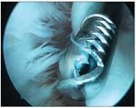
Fraying provides evidence of the internal impingement of the posterior rotator cuff
In addition, a protracted scapula essentially positions the posterior glenoid closer to the humeral head during overhead activities that require abduction. The posterior rotator cuff, particularly the junction of the supraspinatus and the infraspinatus, may become sandwiched between the posterior glenoid and the humeral head during overhead activities in this hyperangulated joint. This "internal impingement," or pinching, of the posterior cuff that some throwers experience will predictably worsen as protraction increases (Figure 1).
A protracted scapula may disturb the natural length-tension relationship of the muscles that are required for optimal function; shoulder weakness is a natural consequence. Again, a scapula that is not firmly stabilized against the thorax cannot effectively serve as a stable platform for both the cuff muscles and the deltoid.1
Athletes who have protracted scapulae may present with variable symptoms. As the scapula "floats" out of position, tension on restraining muscles, such as the levator scapula, may produce posterior shoulder soreness.6 Impingement and anterior shoulder symptoms are common as the supraspinatus is pinched between the less stable humeral head and the encroaching acromion. In addition, posterior joint line pain may manifest as internal impingement symptoms develop.
Origins of protraction
Because the scapula depends on its surrounding musculature for stability, alterations in muscular strength about the shoulder materially affect scapular position. Muscles generally respond to neighboring joint perturbations in 1 of 2 ways: inhibition or contraction. Consider the quadriceps inhibition that manifests rapidly after ligamentous injury to the knee. In contrast, note how hamstring tightness may accompany the onset of an adjacent lumbar disk herniation or stress fracture of the pars interarticularis.
In the periscapular musculature, shoulder derangements tend to inhibit the lower trapezius and serratus; the upper trapezius tends to become more contracted.7-9 The biomechanical consequence of these muscular responses is increased protraction.
Note that by virtue of their origin and insertions, both the lower trapezius and serratus anterior serve as external rotators of the scapula. For example, the serratus anterior inserts on the medial scapula. Weakness or inhibition of this muscle causes prominence of the medial scapular border. Concomitant contracture of the upper trapezius may potentiate inferior medial border prominence. Inhibition of the above-mentioned muscles results in a net internal rotation posture (protraction) of the scapula.
Therefore, the presence of scapular dyskinesis, especially protraction, may indicate that there is a neighboring joint or cuff problem causing reflex inhibition of select neighboring periscapular muscles. For example, a superior labral tear may not only manifest with the mechanical symptoms of catching, locking, and clicking but also demonstrate scapular asymmetry.
Another potential source of protraction in athletes may be related to the shoulder motion alterations that develop in throwers over time. Specifically, a contracture of the posterior inferior shoulder capsule has been shown to develop in many overhead throwers. The origin of this contracture remains speculative; many authors maintain that it is an exuberant capsular healing response to the distractive forces of the "follow-through" phase in the throwing motion. Thomas and associates10 recently demonstrated that the posterior capsule of baseball players is hypertrophied, supporting a fibroblastic healing response to the repetitive microtrauma of throwing.
However, limited internal rotation in abduction is demonstrated in athletes in whom posterior shoulder capsular contracture (glenohumeral internal rotation deficit [GIRD]) develops. For the follow-through to be completed in this position, scapulothoracic motion is increased to accommodate the thrower's need to approximate the arm toward home plate. Specifically, the scapula travels up and around the thorax to afford the necessary internal rotation for follow-through.11 This "scapular windup," as Kibler described it, results in loosening of both static and dynamic soft tissue restraints and an increase in resting scapular protraction. Indeed, Thomas and colleagues12 have shown a correlation between increasing GIRD and scapular asymmetry.
In addition, overuse of shoulder muscles can result in acquired weakness. "Too many innings pitched" may be the cause of the serratus anterior weakness that many throwers exhibit. More frequently, however, disorders of the kinetic chain are responsible for periscapular weakness.
Fractures of the scapula are rare, especially in overhead athletes; they usually occur as high-energy "collision" injuries. However, anatomical changes, such as those seen in a shortened clavicle fracture,13 can disrupt the resting position of the scapula and result in increased protraction.
For example, clavicle fractures that are significantly shortened concomitantly bring the scapula "around" the ellipsoid thorax. The nature of the connections of the scapula to the clavicle, by way of the coracoclavicular ligaments, dictates that clavicular strut shortening will tend to place the scapula in a more internally rotated (protracted) position. In addition, the altered position of the clavicle can cause disruptions of the normal scapular upward rotation and posterior tilting, because the clavicle strut guides scapular motion.
The role of the kinetic chain
The kinetic chain is a series of links that act in coordinated, sequential fashion to generate and amplify a force. As an example, consider "cracking the whip," in which the movement of the whip handle, if brought about properly and smoothly, can translate into significant velocity generation at the whip end. Similarly, the act of throwing beautifully illustrates a biological kinetic chain in which a force is initiated in the lower body and is both translated and amplified via the trunk, shoulder, and upper extremity, culminating in ball release.14
Any disruption in this chain requires the more distal elements to increase their demand and "catch up" to maintain the same velocity. For example, a thrower who has weak hip abductors may demonstrate pelvic instability during windup. An unstable pelvis does not allow for a smooth transfer of force from the lower extremity to the trunk. Because more than 50% of the force generated for the throw originates in the lower extremities, the thrower needs to catch up by increased recruitment of the trunk, shoulder, and upper extremity. The result may be overuse and, therefore, inhibition of periscapular muscles.
A common kinetic chain disruption is seen in GIRD, in which internal rotation of the glenohumeral joint is limited. The scapula is forced to travel up the thorax (scapular windup) to generate internal rotation, and the elbow joint may realize greatly increased demands. Because the humerus is limited in its ability to rotate internally, the elbow is called into increased demand to maintain velocity. Increased demand on the elbow to supply the necessary internal rotation to complete the throw results in increased strain to the ulnar collateral ligament.
Suzuki and associates15 have shown that scapula fatigue leads to increased flexion of the elbow during throwing. Most younger throwers who present with medial elbow pain can be treated successfully by addressing the internal shoulder rotation deficit.
Other proximal kinetic chain abnormalities that may lead to scapular dyskinesis include limitation of lumbar flexion/extension, inadequately rehabilitated ankle sprains, lead leg quad tightness, and internal rotation deficits of a thrower's plant leg. Each of these conditions prompts catch-up distally along the kinetic chain and may translate to overuse of the periscapular muscles or joint injury, causing reflex muscle inhibition.
DIAGNOSIS
Detection of scapular problems
Physical examination of the athlete's shoulder must entail inspection of his or her back. The presence of scapular asymmetry, either when the patient is at rest or during provocative maneuvers, indicates that scapular dyskinesis is present. For the examination, we ask men to remove their shirt; women are best examined wearing a tank top so that both scapulae can be inspected.
Kibler has elegantly described the scapular slide test to detect shoulder dysfunction in athletes. In this test, the athlete's back is inspected with the arms at rest, the hands positioned on the sides of the pelvis, and the arms at 90° of abduction. Asymmetry in scapular position, especially inferior medial or medial scapular prominence, is noted. Because the serratus anterior muscle inserts on the medial border of the scapula, weakness of this muscle manifests as the two most frequently seen dyskinesis presentations, inferior medial and medial scapular prominence (scapular winging).
Resisted eccentric forward flexion may be the most sensitive test to elicit the subtle winging that is seen in serratus anterior weakness. Simply ask the athlete to mildly resist your efforts to lower his arm in a forward-flexed position. In the presence of serratus anterior weakness or inhibition, the inferior medial or medial border of the scapula will become prominent on descent.
In addition, the patient can use 5-lb weights as resistance. The patient is asked to forward flex the shoulder 10 to 12 times, and the examiner watches scapular motion. In a patient with dyskinesis, scapular motion might look normal for the first few repetitions, but the serratus anterior and lower trapezius quickly fatigue and the dyskinetic pattern becomes much clearer.
History and physical examination
Significant structural perturbations of the shoulder, such as significant labral or cuff tears, lead to scapular asymmetry and must be addressed. To discern overuse issues in overhead athletes, obtain a detailed history, including the number of innings pitched or laps swum and how frequently the athlete engages in the activity. Warning signs of overuse in pitchers include more than 100 pitches per week, year-round throwing, and "showcase" events.
A complete biomechanical evaluation should follow, including kinetic chain assessment. Again, pay close attention to potential proximal kinetic chain abnormalities, particularly in throwers. Check lumbar flexibility by asking the athlete to bend over and touch his toes with his knees straight. Look for hip abductor weakness by asking the athlete to perform repeated single-leg deep knee bends. A "dip" of the pelvis with increased valgus of the knee indicates gluteus medius insufficiency, which may result in a proximal breakdown in throwers' kinetic chain. Inadequately rehabilitated ankle sprains usually manifest with diminished ankle dorsiflexion; this is best noted with an athlete's inability to squat fully on the affected limb while keeping the feet flat on the floor.
Internal rotation deficits of the glenohumeral joint should be noted; they are measured only with the scapula stabilized. Burkhart and associates16 have described the "shoulder at risk" as one that demonstrates more than a 25° difference in internal rotation compared with the nonthrowing shoulder.
FIGURE 2


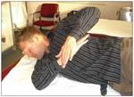
"Sleeper stretches" may be used to address posterior capsular and cuff tightness in overhead athletes
TREATMENT
Posterior capsular and cuff tightness are best addressed by asking athletes to do daily "sleeper" stretches. The patient is asked to lie on his side and perform stretches in 70°, 90°, and 120° of abduction (Figure 2). A stretch should be felt in the posterior shoulder. If pain is felt in the anterior shoulder during stretching, less shoulder abduction should be used until the patient is pain-free. This anterior pain most likely is a replication of impingement symptoms, and it may cause further aggravation of shoulder pain.
Scapular protraction should be mitigated. This is best accomplished with performance of scapular retraction exercises devoted to the muscles most prone to inhibition, the serratus anterior and lower trapezius.
Closed grip rows are an excellent scapular retractor strengthener; the "low row" may be the best exercise for strengthening the serratus anterior. To perform the low row, a "lat bar" is gripped at shoulder level and the athlete pulls the bar slowly to the knees, retracting the scapula at the end of the movement.
Athletes with scapular dyskinesis may have difficulty in performing these exercises correctly because of a lack of neuromuscular control. Patients often are upper trapezius–dominant and perform a shoulder shrug during retraction, which can be counterproductive. Patients may need to use mirrors or be brought through the correct movements manually until they are learned. Verbal cues should be described as "pinching your scapulas down and back."
"Push-ups plus" is another serratus strengthener; the athlete arches his back in an effort to push the body even further than the end position of a conventional push-up. Prone shoulder extension exercises, performed in abduction, address the lower trapezius.
Athletes who have protracted scapulae for several months often experience associated tightening of the pectoralis minor and short head of the biceps. A protracted scapula simply shortens the distance of origin and the insertion of the pectoralis minor; thus, prolonged protraction results in contraction of this muscle. The pectoralis minor is readily stretched by asking the athlete to assume a supine position on a table and placing a rolled towel under the midline of his thorax. Gently push the shoulders toward the table; the contracted pectoralis minor should relax and stretch. Many athletes report this stretch to be not only painless but also "comfortable."
All overhead athletes should be encouraged to engage in a dedicated rotator cuff strengthening regimen that is focused on the infraspinatus, a primary stabilizer for anterior instability. External rotation isotonic exercises that involve use of an elastic band or dumbbells are best performed with the elbow at the side. In this way, the infraspinatus, rather than the deltoid, is isolated. Having the patient initiate the exercise with scapular retraction helps improve both scapular and glenohumeral function. These posterior cuff strengthening exercises should be completed every other day.
Management of scapula-related pain in overhead athletes may be considered successful when the symptoms resolve and the position of the scapula becomes symmetrical with that in the contralateral shoulder. Responses to treatment vary according to symptom severity and duration, but with an early diagnosis, dyskinesis usually resolves with nonoperative approaches. Cases that involve significant "mechanical symptoms," such as locking, catching, or "giving way," probably will require surgical repair.
Resolution of scapula dyskinesis indicates attainment of "joint homeostasis." A scapula returned to its proper position denotes not only abatement of painful stimuli but also restoration of mechanical integrity to the glenohumeral joint.
References:
References
1. Kibler WB, McMullen J. Scapula dyskinesis and its relation to shoulder pain. J Am Acad Orthop Surg. 2003;11:142-151.
2. Warner JJ, Micheli LJ, Arslanian LE, et al. Scapulothoracic motion in normal shoulders and shoulders with glenohumeral instability and impingement syndrome: a study using Moiré topographic analysis. Clin Orthop Relat Res. 1992;285:191-199.
3. Jobe CM, Pink MM, Jobe FW, Shaffer B. Anterior shoulder instability, impingement, and rotator cuff tear: theories and concepts. In: Jobe FW, ed. Operative Techniques in Upper Extremity Sports Injuries. St Louis: Mosby; 1996:164-176.
4. Kibler WB. The role of the scapula in the overhead throwing motion. Contemp Orthop. 1991;22:525-532.
5. Weiser WM. Lee TQ, McMaster WC, McMahon PJ. Effects of scapular protraction on anterior glenohumeral stability. Am J Sports Med. 1999;27:801-805.
6. Burkhart SS, Morgan CD, Kibler WB. The disabled throwing shoulder: spectrum of pathology, part III: the SICK scapula, scapular dyskinesis, the kinetic chain, and rehabilitation. Arthroscopy. 2003;19:641-661.
7. Pink MM, Perry J. Biomechanics of the shoulder. In: Jobe FW, Pink MM, Glousman RE, et al, eds. Operative Techniques in Upper Extremity Sports Injuries. St Louis: Mosby-Year Book; 1996:109-123.
8. McClure PW, Michener LA, Sennett BJ, Karduna AR. Direct 3-dimensional measurement of scapular kinematics during dynamic movements in vivo. J Shoulder Elbow Surg. 2001;10:269-277.
9. McQuade KJ, Dawson J, Smidt GL. Scapulothoracic muscle fatigue associated with alterations in scapulohumeral rhythm kinematics during maximum resistive shoulder elevation. J Orthop Sports Phys Ther. 1998;28:74-80.
10. Thomas SJ, Swanik CB, Higginson JS, et al. A bilateral comparison of posterior capsule thickness and its correlation with glenohumeral range of motion and scapular upward rotation in collegiate baseball players. J Shoulder Elbow Surg. 2011;20:708-716.
11. Fleisig GS, Dillman CJ, Andrews JR. Biomechanics of the shoulder during throwing. In: Andrews JR, Wilk KE, eds. The Athlete's Shoulder. New York: Churchill Livingstone; 1994:355-368.
12. Thomas SJ, Swanik KA, Swanik CB, Kelly JD 4th. Internal rotation deficits affect scapular positioning in baseball players. Clin Orthop Relat Res. 2010;468:1551-1557.
13. Ledger M, Leeks N, Ackland T, Wang A. Short malunions of the clavicle: an anatomic and functional study. J Shoulder Elbow Surg. 2005;14:349-354.
14. Kibler WB. Biomechanical analysis of the shoulder during tennis activities. Clin Sports Med. 1995;14:79-85.
15. Suzuki H, Swanik KA, Huxel KC, et al. The effect of isolated scapular muscle fatigue on shoulder and elbow kinematics. J Sport Rehabil. 2006;15:71-88.
16. Burkhart SS, Morgan CD, Kibler WB. The disabled throwing shoulder: spectrum of pathology, part I: pathoanatomy and biomechanics. Arthroscopy. 2003;19:404-420.


