Prevent patellofemoral pain to prevent knee osteoarthritis?
Persons who have patellofemoral pain (PFP) exhibit higher peak levels of patellofemoral stress than those who do not, according to work conducted at the University of Southern California's Musculoskeletal Biomechanics Research Laboratory (MBRL), and a long-term history of PFP in older adults yields a higher probability that patellofemoral osteoarthritis (OA) will occur.
Persons who have patellofemoral pain (PFP) exhibit higher peak levels of patellofemoral stress than those who do not, according to work conducted at the University of Southern California's Musculoskeletal Biomechanics Research Laboratory (MBRL), and a long-term history of PFP in older adults yields a higher probability that patellofemoral osteoarthritis (OA) will occur. No study to date has defined the relationships among patellofemoral joint stress, pain, and cartilage damage consistent with early-stage arthritis in human subjects, but now MBRL PhD candidate Shawn Farrokhi, DPT, is using advanced imaging techniques and computational musculoskeletal modeling (finite element analysis) to identify the factors that contribute to altered joint stress in persons with PFP.
In acting to transmit forces from the quadriceps muscles to the patellar ligament during knee extension, the patella not only changes the direction of muscular force but also augments the mechanical advantage of the quadriceps by increasing the length of the biomechanical lever about


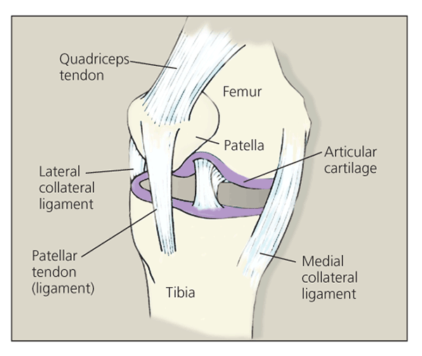
the knee (Figure). The patella also is responsible for force dissipation during knee flexion.
Figure – The patellofemoral region is shown in this drawing. Quantitative MRI analysis of cartilage morphology was used in an experiment to test the hypothesis that persons with patellofemoral pain demonstrate reduced patellar cartilage thickness and volume.
Premature patellofemoral OA-articular cartilage degeneration in the region beneath the kneecap-has long been associated with abnormal patellar alignment and tracking resulting from traumatic or overuse injuries. These injury-based spatial abnormalities are thought to yield unusually high levels of shear and compressive stresses that are the root cause of non–age-related patellofemoral OA.
Painful knee flexion and extension are the primary symptoms of patellofemoral syndrome, which, when experienced long term, is an early marker for patellofemoral joint OA. The pain may result from overloading of the subchondral end plate by forces that the damaged articular cartilage cannot absorb or properly dissipate.
Farrokhi's thesis, "Patellofemoral Joint Stress and Its Relationship to Patellofemoral Joint Pain and Cartilage Changes Consistent With Early Stage Osteoarthritis in Young Adults," includes 2 hypotheses. One is that persons who have PFP exhibit greater peak hydrostatic and octahedral shear stresses than those who are pain-free. The other is that persons with elevated joint stress demonstrate changes in cartilage morphology and composition consistent with early-stage OA (eg, reduced cartilage volume and thickness and elevated resting water content).
To test the first hypothesis, Farrokhi conducted an experiment in which 10 women with PFP and pain-free controls, matched for age, sex, and activity level, underwent MRI knee assessments, 3-dimensional (3D) motion capture, and electromyographic analysis. The resulting anatomical, kinematic, kinetic, and electromuscular data facilitated the creation of weight-bearing–oriented, subject-specific 3D models of knee joint geometry and cartilage morphology. Model-based finite element quasistatic loading simulations were used to quantify the patellofemoral joint cartilage stress profile of each subject while performing a static squatting maneuver.
In a between-group comparison of the amount of stress in model elements representing the patella and femoral chondro-osseous interface, the PFP subjects consistently exhibited higher levels of peak and mean hydrostatic pressure, as well as greater octahedral shear stress, at both 15° and 45° of flexion. The results of an across-group comparison suggested a correlation between higher stress levels and greater degrees of knee flexion.
Quantitative MRI of patellar morphology was used in an experiment designed to test Farrokhi's second hypothesis, that persons with PFP demonstrate reduced patellar cartilage thickness and volume. A secondary objective was to determine whether subjects with PFP tend to exhibit size- or shape-related abnormalities in the bones of the patellofemoral region.
Again, 10 female test subjects were compared with a group of matched pain-free controls. No differences in patellar bone volume or patellar subchondral bone area were evident between subjects with PFP and their matched pain-free counterparts in the MRI data. However, the subjects with PFP demonstrated statistically significant reductions in cartilage volume and thickness.
"The combined finding of elevated hydrostatic pressure and octahedral shear stress across the 2 knee flexion angles supports the premise that PFP may in fact be related to elevated joint stress," Farrokhi noted. This finding, along with those of his second experiment that associate PFP with reductions in cartilage morphology, supports the assertion that patients with PFP may be predisposed to patellofemoral dysfunction because of elevated joint stress. Therefore, treatments aimed at decreasing patellofemoral stresses may be indicated as a prophylactic measure against patellofemoral joint OA in patients who present with PFP.


