A Man With 10 Years of Exquisite Finger Pain
A 25-year-old man was seen in the orthopedic clinic with a complaint of severe, exquisite pain at the ulnar aspect of the distal phalanx of his dominant right index finger. The pain had been present for 10 years, but he had not sought treatment.
A 25-year-old man was seen in the orthopedic clinic with a complaint of severe, exquisite pain at the ulnar aspect of the distal phalanx of his dominant right index finger. The pain had been present for 10 years, but he had not sought treatment. There had been no major injury to his hand. The patient had diet-controlled diabetes mellitus and worked in a job involving computer use. Neither working on the keyboard nor frequently playing tennis produced symptoms. Only direct trauma to the area, such as with tapping directly on the painful spot on his finger, clapping his hands, or throwing a ball would produce marked pain. The patient reported no cold intolerance.


The results of physical examination of the patient’s index finger were remarkable only for marked pain to palpation at the ulnar border of the distal phalanx just adjacent to the base of the fingernail. A slight bluish dot appeared at the ulnar base of the fingernail.
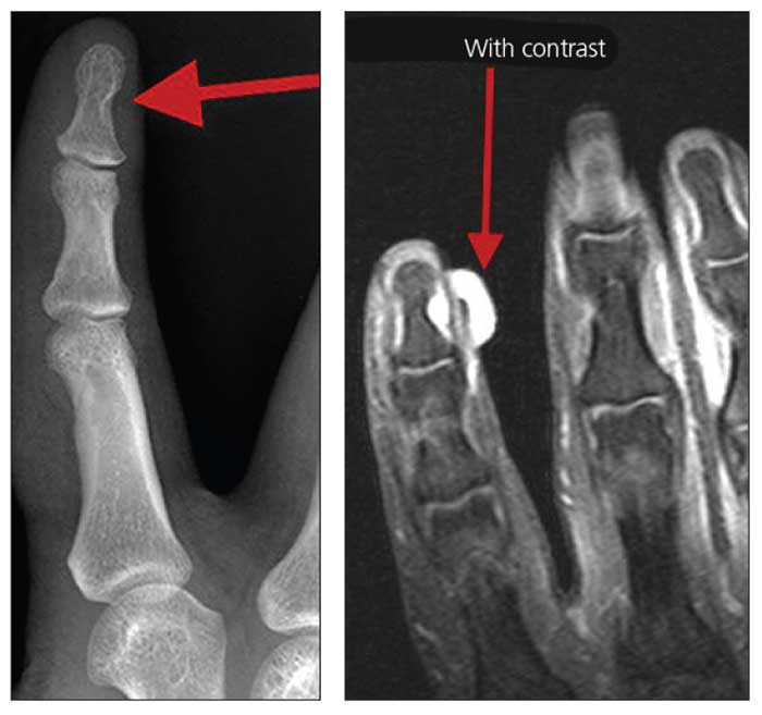
An x-ray film (left) obtained of the patient’s right index finger showed mild chronic scalloping at the ulnar border of the distal phalanx (red arrow). An MRI scan of the same finger, obtained with contrast (right) and without contrast, showed a hyperintense T2 signal, hypointense T1 signal, and intense post-gadolinium enhancement (arrow) corresponding with the area of focal cortical scalloping seen on the x-ray film.
What is your diagnosis?
(Find the answer on the next page.)
The patient underwent surgical excision of a mass from his right index finger under digital block. An intraoperative photograph (below, left) of his right index finger shows a glomus tumor in situ. The specimen was 5 mm long, oval, soft, and tan (below, right).


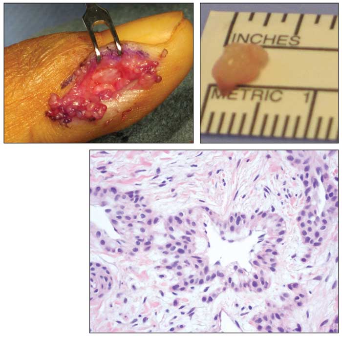
The patient’s pathology report indicated that the mass was consistent with a glomus tumor. Cells with rounded nuclei and eosinophilic cytoplasm were seen in a fibrous stroma with vascular lumina; the photomicrograph (bottom) with hematoxylin and eosin staining shows a vascular channel surrounded by a collarette of glomus cells.
Vascular disorders of the hand are uncommon compared with other problems of the hand, and they are less common than vascular problems found elsewhere in the body.1 Glomus cells are smooth muscle cells that surround small vessels and play a role in the regulation of peripheral blood flow; these tumors, which are benign, account for 1% to 5% of soft tissue tumors of the hand.2
Hand surgeon William Newmeyer, MD, summed up glomus tumors concisely: “Of all the vascular tumors of the upper extremity, this is the one that excites the most interest. The reasons for this are manifold: it is rare, it presents with a striking clinical picture, and when correctly treated, the cure rate is close to 100%.”1
The classic presentation of glomus tumors involves pinpoint finger pain, paroxysmal pain, and hypersensitivity to cold. Cold intolerance is diagnostic when present, but it is not always present in the clinical pattern of these tumors.3
Our patient did not display cold hypersensitivity. In fact, he indicated that on occasion he would plunge his hand into cold water to relieve the intense pain.
Our patient reported a 10-year delay in seeking treatment that he attributed primarily to not realizing that anything could be done about his chronic pain. Many authors have noted protracted delays and misdiagnoses before surgical treatment for glomus tumors is provided.4 The patient made an uneventful recovery after surgery, and his symptoms were relieved.
References:
1. Newmeyer WL. Vascular disorders. In: Green DP, ed. Operative Hand Surgery. 3rd ed. New York: Churchill Livingstone; 1993:2251â2308.
2. Johnston TR, ed. Tumors of the hand and wrist. In: Greene WB, ed. Essentials of Musculoskeletal Care. 2nd ed. Rosemont, IL: American Academy of Orthopaedic Surgeons; 2001:287-289.
3. Lee IJ, Park DH, Park MC, Pae NS. Subungual glomus tumors of the hand: diagnosis and outcome of the transungual approach. J Hand Surgery Eur Vol. 2009;34:685-688.
4. McDermott EM, Weiss AP. Glomus tumors. J Hand Surgery. 2006;31A:1397-1400.


